Tail vein injection of mice with imaging light
Tail vein injection of mice with imaging light
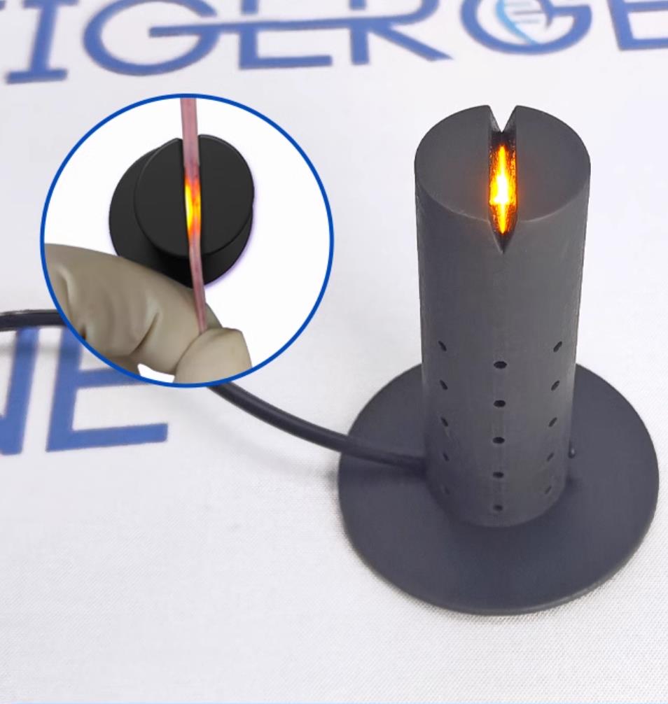
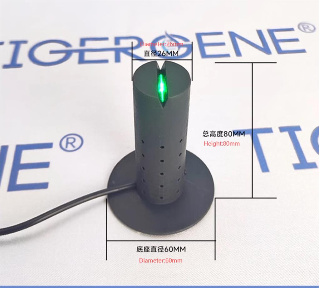
Tail vein injection imaging light, used for the display of veins during tail vein injection in rats and mice.
The device works better in a relatively dark environment, and the light directly above the head can be turned off during use. Some mice have tail veins that are not clearly visible even with the assistance of this device, or they are clearly visible but very small, all due to the fact that the blood vessels themselves too small. The clarity and thickness of the vein image are related to the size and fullness of the blood vessel, so methods such as warming the tail in warm (45°C for 2 minutes), pinching the vein near the heart, and rubbing the tail against the fur can dilate the tail veins. The tail veins mice are very superficial (about 0.3MM under the skin), and the needle should be inserted with the bevel facing up and almost parallel to the tailIt is necessary to master the basic operation skills of tail vein injection and to adapt to this device through a certain number of practices. It is recommended to use a 0G needle with a 1ml syringe or an insulin needle for tail vein injection in mice.
无法加载取货服务可用情况
98 件存货
查看完整详细信息


